水母伤口几分钟愈合:科学家欲破解其无疤修复机制
本文来自海洋生物实验室(MBL),报道了芝加哥大学副教授 Jocelyn Malamy 对透明水母 Clytia hemisphaerica 伤口愈合机制的最新研究。水母的伤口愈合速度极快——小伤口几分钟内闭合,大伤口不到一小时,且不留疤痕。其透明身体让研究人员能在活体中实时观察上皮细胞的修复过程。Malamy 在《Molecular Biology of the Cell》上发表的新论文指出,所有上皮伤口愈合均由两种关键细胞结构顺序驱动:首先,伤缘细胞伸出富含肌动蛋白的片状伪足(lamellipodia)像“足状触角”一样爬过基底膜,拖动细胞前移;随后,在伪足后部形成肌动球蛋白环(actomyosin cable),像“荷包绳”一样将伤口拉拢。对于基底膜受损的大伤口,整个上皮片层还会发生集体迁移。该发现统一了此前关于不同生物、不同大小和形状伤口愈合机制的争论。文章适合对发育生物学、再生医学和细胞力学感兴趣的工程师。</summary>
A decade ago this summer, at the Marine Biological Laboratory, Jocelyn Malamy watched jellyfish cells walk toward each other to close a wound for the first time. An associate professor in Molecular Genetics and Cell Biology at the University of Chicago, Malamy had received transparent, dime-sized medusae of the species Clytia hemisphaerica from Evelyn Houliston's lab at the Marine Observatoire in Villefranche.
十年前的那个夏天,在海洋生物学实验室,乔斯林·马拉米第一次亲眼目睹水母细胞“行走”着相互靠拢,闭合伤口。这位芝加哥大学分子遗传学与细胞生物学副教授,从维勒弗朗什海洋观测站的伊芙琳·霍利斯顿实验室获得了透明、一角硬币大小的半球美螅水母。
The medusa, the free-swimming form most people picture when they hear the term jellyfish, is only one stage of the animal's life cycle. At some point in their life cycle, the colony will start releasing baby medusae. In this way, Clytia's medusae forms are much like flowers, while the polyps are like the shrubs that produce them. We tend to think of the flower—or the jellyfish—as the organism, but these are actually reproductive units. The primary form is the shrub or, in Clytia's case, the polyp colony. Like flowers, medusae are short-lived, surviving only a few months at most. The polyp colonies that produce them, however, can persist indefinitely, much like a perennial shrub that blooms year after year.
人们听到“水母”一词时想到的通常是自由游动的水母体,但这只是它们生命周期的一个阶段。马拉米说:“半球美螅水母属于水螅纲,所以大部分时间它们以水螅体群落的形式存在,附着在岩石表面、码头或水下叶片背面。”在生命周期的某个节点,水螅群体会释放幼年水母体。这样看来,水母体就像花朵,而水螅体则是开出花朵的灌木。我们往往把花——也就是水母——当作有机体本身,但水母实际上只是繁殖单元。其主体形式是灌木,对半球美螅水母而言就是水螅体群落。像花朵一样,水母体寿命很短,最多只能存活几个月。而孕育它们的水螅体群落却能无限延续,就像年年盛开的多年生灌木。
Yet unlike other organisms, Clytia medusae can repair damage so rapidly you can actually watch small wounds close within minutes. Larger wounds heal in less than an hour, a rate of recovery humans can only dream of. And unlike in humans, no scar tissue is formed. Instead, Malamy says healing in the jellyfish looks more like embryonic healing, which is scar-free.
但与其它生物不同的是,半球美螅水母的损伤修复速度快到你可以亲眼看着小伤口在几分钟内闭合。较大的伤口也能在一个小时内愈合——这种恢复速度是人类做梦都想要的。而且,与人类不同,这些伤口不会形成疤痕组织。相反,马拉米说:“水母的愈合更像胚胎期的无瘢痕愈合。”
These traits make Clytia a unique window into wound healing. The medusae are transparent, allowing researchers to watch cells move in live animals in real time. Their wounds heal rapidly, and unlike mammals there is no immune system to trigger inflammation around a wound or capillary regeneration to obscure the basic mechanics of repair. As a result, scientists can observe epithelial cells stitching damaged tissue back together. Even more importantly, many of the fundamental mechanisms of wound healing appear to be conserved. A lot of the processes that we see in Clytia's wound healing are really similar to what you see in all other systems, including mammalian systems, Malamy explains. In fact, she adds When you're staring at these epithelial cells, you wouldn't know this was a jellyfish. It could be any kind of squamous epithelial cell sheet, and that's nice, because it means that hopefully what we learn in jellyfish can give us insights into other animals as well.
这些特性使半球美螅水母成为研究伤口愈合的独特窗口。水母体是透明的,研究人员可以在活体动物身上实时观察细胞运动。它们的伤口愈合迅速,而且与哺乳动物不同,没有免疫系统触发伤口周围的炎症反应,也没有毛细血管再生来干扰修复的基本机制。因此,科学家可以清楚地看到上皮细胞如何将受损组织缝合起来。更重要的是,许多伤口愈合的基本机制似乎是保守的。马拉米解释说:“我们在半球美螅水母伤口愈合中看到的很多过程,和你在所有其他系统——包括哺乳动物系统——中看到的非常相似。”她补充说:“当你盯着这些上皮细胞时,你根本看不出这是水母。它可以是任何一种鳞状上皮细胞片,这很好,因为这意味着我们在水母身上学到的东西有望为其它动物提供洞见。”
Epithelial cells cover all the body's surfaces. They make up our skin and line the inside of tissues like the gut. Because both the skin and internal epithelial tissues are regularly damaged and must repair themselves, they are a key focus of wound-healing research. Malamy first characterized Clytia epithelial wound healing in 2017 in work initiated with a group of students while she was a Whitman Fellow at the Marine Biological Laboratory (MBL), and she expanded on that work in a paper she co-authored with MBL faculty member Michael Shribak in 2018. Malamy's new paper in the journal Molecular Biology of the Cell uses Clytia to tackle the confusion in the field over the various mechanisms that have been shown to heal epithelial wounds in different organisms, and wounds of different sizes and shapes. She shows that all epithelial wound healing in Clytia is driven by two key cellular structures that act in sequence to close a wound, and defines a mechanism that explains how these structures are coordinated in all types of wounds.
上皮细胞覆盖了身体的所有表面。它们构成了我们的皮肤,并铺衬在肠道等组织内部。由于皮肤和内部上皮组织经常受损并需要自我修复,它们成为伤口愈合研究的核心焦点。马拉米2017年首次描述了半球美螅水母的上皮伤口愈合,当时她作为惠特曼研究员在海洋生物学实验室(MBL)与一组学生启动了这项研究;2018年她与MBL教员迈克尔·什里巴克合著论文,进一步拓展了该工作。马拉米在《分子生物学杂志》上发表的新论文,利用半球美螅水母解决了该领域长期以来对在不同生物体中、以及不同大小和形状伤口中愈合的各种机制所存在的困惑。她表明,半球美螅水母的所有上皮伤口愈合都由两种关键细胞结构依次作用来闭合伤口,并定义了一个机制,解释了这些结构在所有类型伤口中是如何协调的。
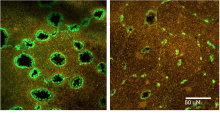
The first structures to form in response to a wound are lamellipodia, which Malamy describes as foot-like feelers that are actin-rich extensions of the cell. These feelers act as explorers and have an almost fluid-like motion, similar to amoebas. Lamellipodia extend out of cells at the edge of wounds and crawl across the basement membrane, a protein sheet that's underneath all epithelial cells in all systems, she explains. As they walk, they drag the cells that produced them forward, eventually stretching the cell body over a wound to close it. Malamy shows in her new paper that these lamellipodia form even in tiny wounds internal to a single cell, a novel observation.
伤口结成的第一时间形成的结构是片状伪足(lamellipodia),马拉米将其描述为“足状的触须,是细胞富含肌动蛋白的延伸部分”。这些触须充当探索者,运动起来几乎像流体,类似于变形虫。她解释说,片状伪足从伤口边缘的细胞中伸出,在基底膜——一种位于所有系统上皮细胞下方的蛋白质片层——上爬行。当它们“行走”时,拖着产生它们的细胞前进,最终将细胞体拉伸覆盖伤口,使其闭合。马拉米在新论文中表明,即使是在单个细胞内部形成的微小伤口中,也会形成这些片状伪足,这是一个新的发现。
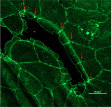
As the lamellipodia crawl forward, a second wound-healing mechanism comes into play: an actomyosin cable forms at the back of the lamellipodia as they walk forward. As soon as the lamellipodia cover the basement membrane, the cable is triggered to contract. An actomyosin purse string pulls cells across a region where the basement membrane is damaged. A microscopy video shows this process.
随着片状伪足向前爬行,第二种伤口愈合机制开始发挥作用:在片状伪足向前移动的过程中,其后方会形成一个肌动球蛋白环。一旦片状伪足覆盖了基底膜,这个环就会被触发收缩。就像一个“肌动球蛋白的钱包吊绳”,它将细胞拉过基底膜受损的区域。一段显微视频展示了这一过程。
An actomyosin cable structure is also particularly important if the basement membrane has been damaged. If the lamellipodia have run into some debris or a tear in the basement membrane, and they can't go any further, Malamy says the actin cable can pull the cells over the basement membrane damage and also expel wound debris. As long as there is a basement membrane, lamellipodia will advance, but if the wound is too big, no matter how much the lamellipodia stretch their cells, they won't reach each other. This leads to a collective cell migration being triggered, where the entire sheet of epithelium lifts itself up and starts walking, Malamy explains. Once the lamellipodia of the cells in the front meet, this large wound can then close the same way as smaller wounds.
当基底膜受损时,肌动球蛋白环结构也特别重要。马拉米说:“如果片状伪足撞到了碎片或基底膜的撕裂处,无法继续前进,肌动球蛋白环可以把细胞拉过基底膜损伤区域,同时还能排出伤口碎片。” 只要有基底膜在,片状伪足就会继续推进;但如果伤口太大,无论片状伪足如何拉伸细胞,它们都无法彼此接触。这就会触发集体细胞迁移——整个上皮片层“自行抬高并开始行走”,马拉米解释道。一旦前面细胞的片状伪足相遇,这个大伤口就能以与小伤口相同的方式闭合。
This is a truly elegant mechanism where the system can rapidly adapt to heal all the kinds of wounds that might occur in nature Malamy says.
“这是一个真正优雅的机制,系统可以快速适应,愈合自然界可能出现的各种伤口,”马拉米说。
Malamy is next planning to examine the mechanisms driving basement membrane repair. It's great that you can heal a wound by dragging the cells over it, she explains, but at some point, a damaged basement membrane has to get fixed. She hopes to find out how that happens in Clytia as it's currently unclear how basement membrane repair happens in any system.
马拉米下一步计划研究驱动基底膜修复的机制。她解释说:“能把细胞拖过去愈合伤口固然很好,但在某个时刻,受损的基底膜必须得到修复。”她希望弄清楚这个过程在半球美螅水母中是如何发生的,因为目前还不知道基底膜修复在任何系统中是如何进行的。